3D Ultrasound Atlas
Comparative fetal anatomy
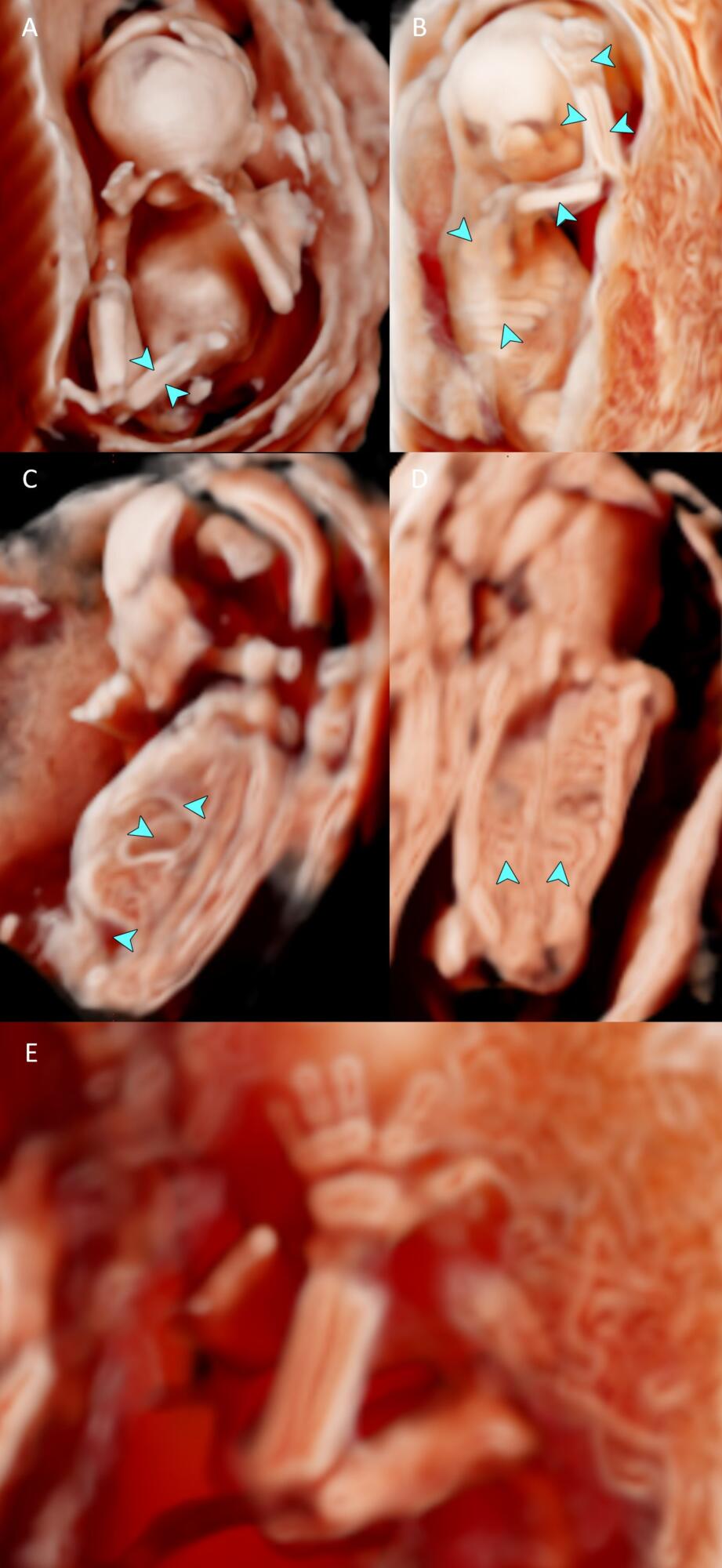
Below are annotated images of a fetus at 17 weeks gestation, imaged in-vivo using 3D ultrasound imaging with CrystalVue™ and RealisticVue™ applied.

A and B – fetal skeleton including long and short bones, ribs and scapula at 17 weeks gestation. C – sagittal view of the diaphragm and hypoechoic stomach and bladder at 17 weeks gestation. D – coronal view of fetal kidneys at 17 weeks gestation. E – metacarpals, radius and ulna


