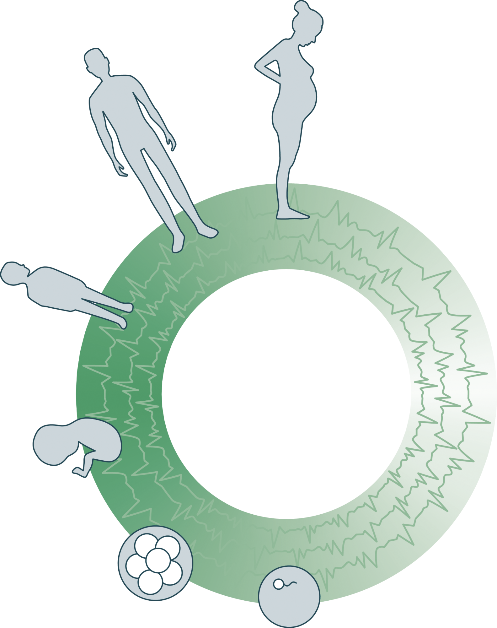
“Our real teacher has been and still is the embryo, who is, incidentally, the only teacher who is always right.”
– Viktor Hamburger (1900-2001) –
The 3D Atlas of Human Embryology comprises 14 user-friendly and interactive 3D-PDFs of all organ systems in real human embryos between Carnegie stages 7 and 23 (15 till 60 days of development), and additional stacks of digital images of the original histological sections and annotated digital label files.
The atlas was created by students and embryologists of the Department of Anatomy, Embryology & Physiology of the Academic Medical Center (AMC) in Amsterdam, the Netherlands and it is made freely available to the scientific community to facilitate veracious embryology education and research.
Click here to access the Science publication, and feel free to donate to our research.
”Unlocking the potential of state-of-the-art 3D ultrasound technology for early fetal anatomy assessment
3D Ultrasound Atlas


