A Dream Realized: 3D Real-Time Embryology is Here!
8-09-2024
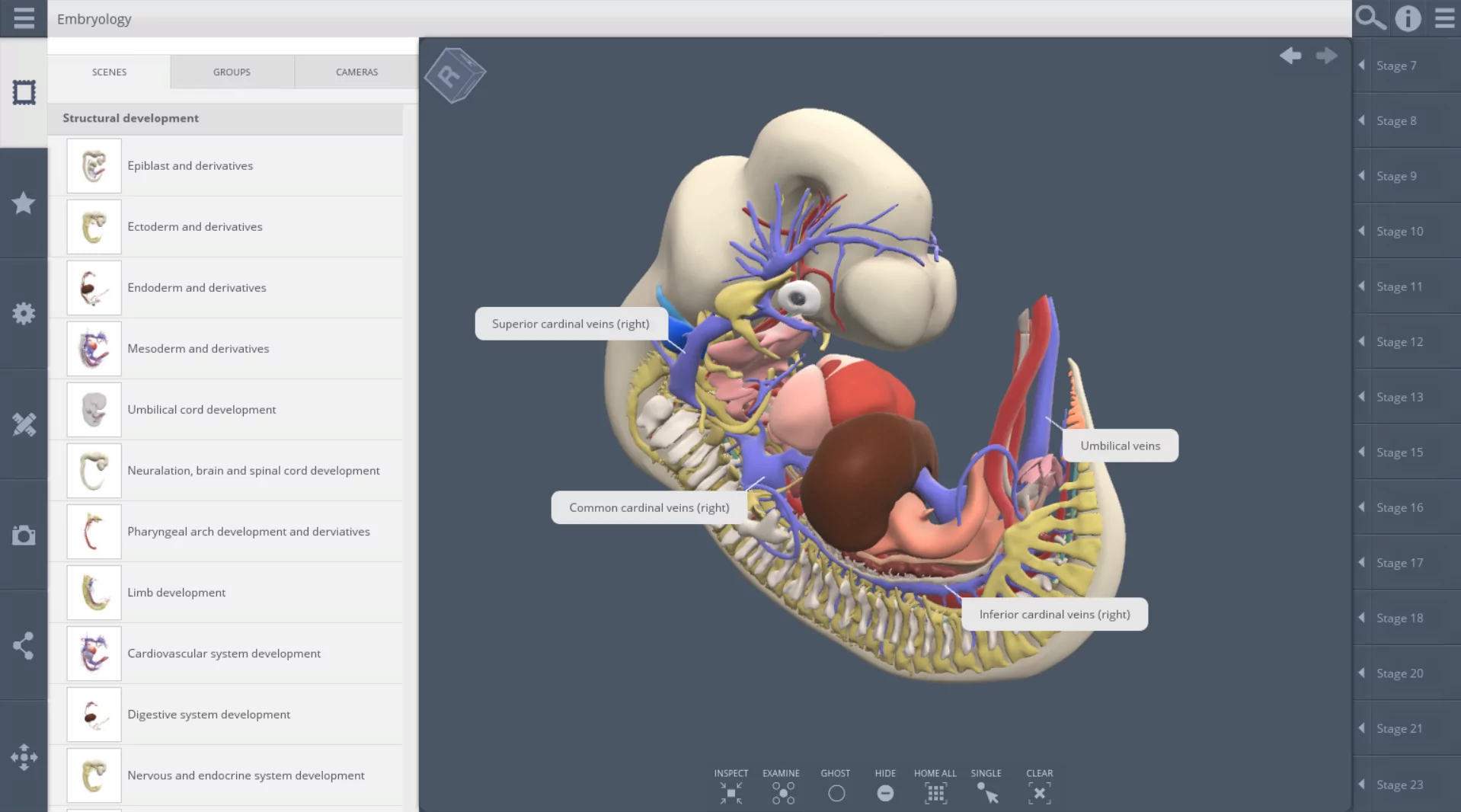
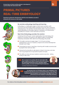
After years of dedication and collaboration, the groundbreaking vision of transforming the 3D Embryo Atlas into a comprehensive digital educational resource has finally become a reality. Originally published in Science in 2016, this atlas has now evolved into an accessible, real-time 3D learning tool, thanks to the exceptional efforts of Primal Pictures and their talented team of anatomists, including Lorna Wilson and Naomi Senior, alongside their skilled graphic designers.
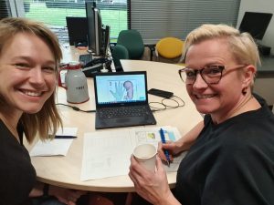
Dr. Bernadette de Bakker and Lorna Wilson of Primal Pictures sketching the first ideas for the atlas in 2022
Embryology, often considered one of the most challenging subjects in medical education, has long been a focus for making complex concepts easier to understand for both students and clinicians. The launch of this innovative embryology application, now integrated into Primal Pictures’ renowned digital anatomy atlas, marks a significant step forward in educational tools for the medical field.
This project would not have been possible without the contributions of over 80 students who played a crucial role in the development of the 3D Embryology Atlas. Additionally, the support of key colleagues and team members was instrumental in bringing this vision to life. Special recognition goes to Marc Roelofs, Steven van Huiden, and Joris Heus from IXA – Innovation Exchange Amsterdam, Manja van Riet-Colmans of AMR, and Rik Bouman from the Legal department, whose efforts greatly facilitated this collaboration.
With the launch of this 3D Real-Time Embryology tool, educators and learners can look forward to a new era of accessible, engaging, and effective embryology education. This milestone not only enhances the learning experience but also reinforces the importance of interdisciplinary collaboration in advancing medical education.
To learn more about the launch of the 3D Real-Time Embryology tool, visit Primal Pictures’ announcement.
This partnership with Primal Pictures is a great example of how Amsterdam UMC continues to lead the way in innovative medical education.
The Cold Spring Harbor Asia (CSHA) Meeting on Human Development
Read moreAUF Impact Award for citizen science
We are excited to share that we have been awarded the Impact Award from the Amsterdam University Fun…
Read moreDaphne Naessens receives two grants for research on brain cleansing
Daphne Naessens receives two grants for research on brain cleansing We are pleased to announce that…
Read more
