Unlocking the shape of the fetal heart
1-10-2023
Unlocking the shape of the fetal heart: a collaborative research initiative
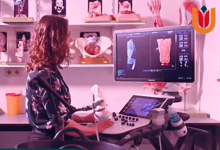
Last September, PhD student Marieke Buijtendijk visited the Perinatal Imaging Department at King’s College Hospital in London to join forces with an highly esteemed research group led by David Lloyd and Maria Perez. Thanks AR&D’s financial support, she was able to embark on a one-month work visit to kick-start this promising collaborative research project.
Our goal: bringing AI to fetal cardiac ultrasound imaging
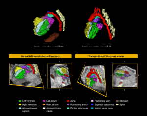
Our ultimate goal is to use artificial intelligence to generate three-dimensional (3D) models of the fetal heart, offering an in-depth exploration of its 3D shape through ultrasound volumes. While our collaborators had previously achieved this with fetal cardiac MRI, the transition to ultrasound posed unique challenges that warranted attention before to make further analyses possible.
Navigating ultrasound challenges
Tackling these challenges involved developing a method to align ultrasound volumes consistently. By manually selecting anatomical landmarks and employing several processing steps, we successfully generated an averaged 3D ultrasound atlas of normal fetal hearts. This atlas serves as a valuable research tool, streamlining future studies by making automated alignment of ultrasound volumes possible and unlocking diverse research possibilities.
Marieke is immensely grateful to AR&D for funding this visit, which broadened her insights into using 3D fetal cardiac imaging data, which she can apply to her PhD research. Our ultimate mission is to deepen our understanding of normal and abnormal fetal cardiac anatomy, a crucial step in advancing prenatal screening for congenital heart disease.
Publication: Early thyroid development in Down syndrome
We are pleased to share a new publication in Human Molecular Genetics that sheds light on why thyroi…
Read moreA Milestone: 100 Scientific Publications!
I’m proud to share my 100th scientific publication: a review paper on an incredibly important yet st…
Read moreThe 4D Embryonic Brain Atlas
Dr. de Bakker and colleagues have published the 4D Human Embryonic Brain Atlas (8–12 weeks), a spati…
Read more